2021
|
Kinch MS, Kraft Z, Schwartz T Drug Discov Today. 2021 Jan;26(1):240-247. doi: 10.1016/j.drudis.2020.10.026. The biopharmaceutical industry has undergone remarkable changes over the past half century, driven largely by a need to offset the ever-rising costs of developing new medicines. In this report, we aggregated information about the creation and fate of all clinical-stage biopharmaceutical companies, assessing trends over time. These results reveal that the rate of new company formation has been declining at the same time that industry consolidation has been accelerating at an unprecedented rate. Consequently, the number of companies involved in biopharmaceutical research and development has declined by one-third over the past decade, while those able to achieve at least one FDA approval has dropped by more than half. These findings raise important questions about the sustainability of an industry that is vital for both public and economic health. |
|
Kinch MS, Kraft Z, Schwartz T Drug Discov Today. 2021 https://doi.org/10.1016/j.drudis.2021.07.003 Amid a global pandemic, the US Food and Drug Administration (FDA) remained relatively active, approving 55novel molecular entities (NMEs) in 2020, the third highest annual rate recorded. Orphan approvals also surged, capturing 60% of NMEs introduced during 2020, as did the number of NMEs approved using a priority review. The pandemic did appear to impact one recent trend, and in a paradoxically encouraging way. Escalating rates of consolidation slowed in 2020, with only 102 companies lost, down by two-thirds over the rate in 2019. This leaves 2000 extant clinical-stage pharmaceutical companies. When limiting this analysis to companies contributing to the research and development (R&D) of an approved drug, eight were lost, leaving 144 extant. |
2020
|
Dang, X., Zhang, L., Franco, A., Li, J., Rocha, A. G., Devanathan, S., Dolle, R. E., Bernstein, P. R., & Dorn, G. W., 2nd J. Med. Chem. 2020, 63, 13, 7033–7051 Mutations in the mitochondrial fusion protein mitofusin (MFN) 2 cause the chronic neurodegenerative condition Charcot-Marie-Tooth disease type 2A (CMT2A), for which there is currently no treatment. Small-molecule activators of MFN1 and MFN2 enhance mitochondrial fusion and offer promise as therapy for this condition, but prototype compounds have poor pharmacokinetic properties. Herein, we describe a rational design of a series of 6-phenylhexanamide derivatives whose pharmacokinetic optimization yielded a 4-hydroxycyclohexyl analogue, 13, with the potency, selectivity, and oral bioavailability of a preclinical candidate. Studies of 13cis– and trans-4-hydroxycyclohexyl isostereomers unexpectedly revealed functionality and protein engagement exclusively for the trans form, 13B. Preclinical absorption, distribution, metabolism, and excretion (ADME) and in vivo target engagement studies of 13B support further development of 6-phenylhexanamide derivatives as therapeutic agents for human CMT2A. |
|
B. Case, P. W.Rothlauf, R. E. Chen, Z. Liu, H. Zhao, A. S. Kim, L.-M. Bloyet, Q. Zeng, S. Tahan, L. Droit, M.X.G.Ilagan, M. A. Tartell, G. Amarasinghe, J.P. Henderson, S. Miersch, M. Ustav, S. Sidhu, H.W.Virgin, D. Wang, S. Ding, D. Corti, e.S. Theel, D.H.Fremont, M.S. Diamond and S.P.J.Whelan Cell Host & Microbe. Volume 28, Issue 3, 9 September 2020, Pages 475-485.e5 Antibody-based interventions against SARS-CoV-2 could limit morbidity, mortality, and possibly transmission. An anticipated correlate of such countermeasures is the level of neutralizing antibodies against the SARS-CoV-2 spike protein, which engages with host ACE2 receptor for entry. Using an infectious molecular clone of vesicular stomatitis virus (VSV) expressing eGFP as a marker of infection, we replaced the glycoprotein gene (G) with the spike protein of SARS-CoV-2 (VSV-eGFP-SARS-CoV-2) and developed a high-throughput-imaging-based neutralization assay at biosafety level 2. We also developed a focus-reduction neutralization test with a clinical isolate of SARS-CoV-2 at biosafety level 3. Comparing the neutralizing activities of various antibodies and ACE2-Fc soluble decoy protein in both assays revealed a high degree of concordance. These assays will help define correlates of protection for antibody-based countermeasures and vaccines against SARS-CoV-2. Additionally, replication-competent VSV-eGFP-SARS-CoV-2 provides a tool for testing inhibitors of SARS-CoV-2 mediated entry under reduced biosafety containment.
|
|
Rising Academic Contributions to Drug Development: Evidence of Vigor or Trauma? Kinch MS, Horn C, Kraft Z, Schwartz T ACS Pharmacol. Transl. Sci. 2020, 3, 6, 1427–1429 We analyzed therapeutic areas most commonly targeted by academia since 2001, finding a domination of certain oncology and infectious diseases. These findings raise important questions about whether this trend reflects an expanded opportunity arising from academic research or a troubling sign of an industry struggling with the challenges of innovation. |
|
Kinch, MS, Henderson, JP. Scientific American. https://www.scientificamerican.com/article/how-politics-muddied-the-waters-on-a-promising-covid-19-treatment/ On Sunday afternoon, August 24, President Trump announced an emergency use authorization (EUA) for what he described as a “powerful therapy” against COVID-19. Known as convalescent plasma, or CP, it’s donated by survivors of the disease, on the theory that the antibodies it carries can protect others from COVID’s worst ravages. And a few weeks ago, the Mayo Clinic released a preliminary report saying CP showed promise in doing just that—but nevertheless, the New York Times reported that the FDA was not ready to approve the treatment, even on an emergency basis. |
|
Kinch MS, Horn, C, Kraft Z, Schwartz T. Drug Discovery Today, Sep 11:S1359-6446(20)30342-1. doi: 10.1016/j.drudis.2020.09.004. An assessment of inventors of US Food and Drug Administration (FDA)-approved medicines reveals a growing role for academic entrepreneurship in general and National Institutes of Health (NIH)-supported investigators in particular. For all small-molecule therapeutics approved between 2001 and 2019 (383 in total), 8.3% listed an academic inventor in the Orange Book. Remarkably, an additional 23.8% listed an inventor from a company founded by an NIH-funded academic inventor. Over time, the relative inventive contributions from academia has progressively increased, including nearly one-third of medicines approved since 2017. These findings suggest a surging role for academic inventors and founders, perhaps in combination with a faltering of traditional private sector dominance of drug discovery. |
|
Kinch MS ACS Pharmacology & Translational Sciences, https://doi.org/10.1021/acsptsci.0c00138. The COVID-19 pandemic has been humbling for the biomedical community, pointing out as much about what we do not know as what we do. Among these learnings are lessons about immune-based measures to prevent or treat a new biothreat. This article summarizes lessons learned from two experimental approaches for passive immunity, convalescent plasma and monoclonal antibody therapy. Two early reports of outcomes, both of which appeared within hours of one another, reveal the importance of blending past learning with a forward-looking approach. These also present cautionary lessons as the world looks to new vaccines to help eradicate this deadly scourge. |
|
Kinch MS Drug Discov Today 2020 Sep 11:S1359-6446(20)30341-X. doi: 10.1016/j.drudis.2020.09.002. The US Food and Drug Administration (FDA) green-lighted the marketing of 53 therapeutic agents in 2019. This rate of approvals was consistent with the 5-year running average. Nonetheless, a few changes are worth noting. The rate of medicines first approved using an orphan drug designation declined from 56% in 2018 to 41% in 2019, which mirrored a comparable decline in the use of priority review. A second notable feature was an uptick in industry consolidation. Twenty-five companies were lost, primarily because of mergers, leaving only 146 extant companies that have contributed to the research or development of an innovative FDA-approved medicine. |
2019
|
Damalanka, V.C., Han, Z., Karmakar, P., O’Donoghue, A.J., La Greca, F., Kim, T., Pant, S.M., Helander, J., Klefstrom, J., Craik, C.S., et al. J. Med. Chem. 2019, 62, 2, 480–490 Matriptase and hepsin belong to the family of type II transmembrane serine proteases (TTSPs). Increased activity of these and the plasma protease, hepatocyte growth factor activator (HGFA), is associated with unregulated cell signaling and tumor progression through increased MET and RON kinase signaling pathways. These proteases are highly expressed in multiple solid tumors and hematological malignancies. Herein, we detail the synthesis and structure–activity relationships (SAR) of a dipeptide library bearing Arg α-ketobenozothiazole (kbt) warheads as novel inhibitors of HGFA, matriptase, and hepsin. We elucidated the substrate specificity for HGFA using positional scanning of substrate combinatorial libraries (PS-SCL), which was used to discover selective inhibitors of matriptase and hepsin. Using these selective inhibitors, we have clarified the specific role of hepsin in maintaining epithelial cell membrane integrity, known to be lost in breast cancer progression. These selective compounds are useful as chemical biology tools and for future drug discovery efforts. |
|
Up to 15% of couples experience infertility. Implantation must occur in order for the pregnancy hormone hCG to be detected, and thus many pregnancies go unrecognized before being lost due to implantation failure. Decidualization is a complex differentiation process during which the uterine lining grows and dramatically changes form in response to ovarian hormone stimulus, and this process is required for successful implantation. By developing and utilizing a new reporter cell line, the present study systematically uncovers more than 4,000 genetic and chemical modulators of the human decidual response in the hopes of catalyzing new treatments for female infertility, subfertility, and recurrent pregnancy loss. |
|
Griesenauer RH, Schillebeeckx C, Kinch MS Database, doi: 10.1093/database/baz087 The Clinical Drug Experience Knowledgebase (CDEK) is a database and web platform of active pharmaceutical ingredients with evidence of clinical testing as well as the organizations involved in their research and development. CDEK was curated by disambiguating intervention and organization names from ClinicalTrials.gov and cross-referencing these entries with other prominent drug databases. Approximately 43% of active pharmaceutical ingredients in the CDEK database were sourced from ClinicalTrials.gov and cannot be found in any other prominent compound-oriented database. The contents of CDEK are structured around three pillars: active pharmaceutical ingredients (n = 22 292), clinical trials (n = 127 223) and organizations (n = 24 728). The envisioned use of the CDEK is to support the investigation of many aspects of drug development, including discovery, repurposing opportunities, chemo- and bio-informatics, clinical and translational research and regulatory sciences. |
|
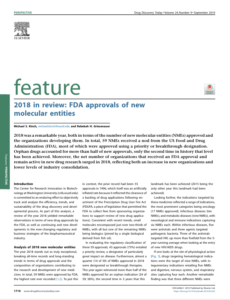
Griesenauer RH, Kinch MS. Drug Discov Today. 2019 May 31. pii: S1359-6446(19)30114-X. doi: 10.1016/j.drudis.2019.05.022 2018 was a remarkable year, both in terms of the number of new molecular entities (NMEs) approved and the organizations developing them. In total, 59 NMEs received a nod from the US Food and Drug Administration (FDA), most of which were approved using a priority or breakthrough designation. Orphan drugs accounted for more than half of new approvals, only the second time in history that level has been achieved. Moreover, the net number of organizations that received an FDA approval and remain active in new drug research surged in 2018, reflecting both an increase in new organizations and lower levels of industry consolidation. |
|
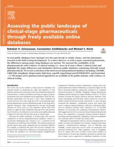
Griesenauer RH, Schillebeeckx C, Kinch MS. Drug Discov Today 2019 Jan 25. pii: S1359-6446(18)30258-7. doi: 10.1016/j.drudis.2019.01.010 Several public databases have emerged over the past decade to enable chemo- and bio-informatics research in the field of drug development. To a naive observer, as well as many seasoned professionals, the differences among many drug databases are unclear. We assessed the availability of all pharmaceuticals with evidence of clinical testing (i.e., been in at least a Phase I clinical trial) and highlight the major differences and similarities between public databases containing clinically tested pharmaceuticals. We review a selection of the most recent and prominent databases including: ChEMBL, CRIB NME, DrugBank, DrugCentral, PubChem, repoDB, SuperDrug2 and WITHDRAWN, and found that ∼11700 unique active pharmaceutical ingredients are available in the public domain, with evidence of clinical testing. |
|
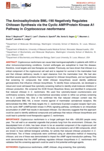
Maybruck, B.T., W.C. Lam, C.A. Specht, M.X.G. Ilagan, M.J. Donlin, and J.K. Lodge. Cryptococcus neoformans can cause fatal meningoencephalitis in patients with AIDS or other immunocompromising conditions. Current antifungals are suboptimal to treat this disease; therefore, novel targets and new therapies are needed. Previously, we have shown that chitosan is a critical component of the cryptococcal cell wall and is required for survival in the mammalian host and that chitosan deficiency results in rapid clearance from the mammalian host. We had also identified several specific proteins that were required for chitosan biosynthesis, and we hypothesize that screening for compounds that inhibit chitosan biosynthesis would identify additional genes/proteins that influence chitosan biosynthesis. To identify these compounds, we developed a robust and novel cell-based flow cytometry screening method to identify small-molecule inhibitors of chitosan production. We screened the ICCB Known Bioactives library and identified 8 compounds that reduced chitosan in C. neoformans. We used flow cytometry-based counterscreens and confirmatory screens, followed by a biochemical secondary screen to refine our primary screening hits to 2 confirmed hits. One of the confirmed hits that reduced chitosan content was the aminoalkylindole BML-190, a known inverse agonist of mammalian cannabinoid receptors. We demonstrated that BML-190 likely targets the C. neoformans G-protein-coupled receptor Gpr4 and, via the cyclic AMP (cAMP)/protein kinase A (PKA) signaling pathway, contributes to an intracellular accumulation of cAMP that results in decreased chitosan. Our discovery suggests that this approach could be used to identify additional compounds and pathways that reduce chitosan biosynthesis and could lead to potential novel therapeutics against C. neoformans. . |
|
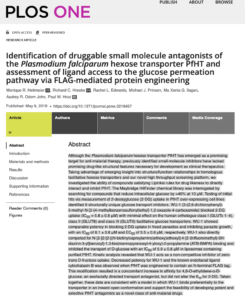
Heitmeier MR, Hresko RC, Edwards RL, Prinsen MJ, Ilagan MXG, Odom John AR, Hruz PW. PLoS One. https://doi.org/10.1371/journal.pone.0216457 Although the Plasmodium falciparum hexose transporter PfHT has emerged as a promising target for anti-malarial therapy, previously identified small-molecule inhibitors have lacked promising drug-like structural features necessary for development as clinical therapeutics. Taking advantage of emerging insight into structure/function relationships in homologous facilitative hexose transporters and our novel high throughput screening platform, we investigated the ability of compounds satisfying Lipinksi rules for drug likeness to directly interact and inhibit PfHT. The Maybridge HitFinder chemical library was interrogated by searching for compounds that reduce intracellular glucose by >40% at 10 μM. Testing of initial hits via measurement of 2-deoxyglucose (2-DG) uptake in PfHT over-expressing cell lines identified 6 structurally unique glucose transport inhibitors. WU-1 (3-(2,6-dichlorophenyl)-5-methyl-N-[2-(4-methylbenzenesulfonyl)ethyl]-1,2-oxazole-4-carboxamide) blocked 2-DG uptake (IC50 = 5.8 ± 0.6 μM) with minimal effect on the human orthologue class I (GLUTs 1–4), class II (GLUT8) and class III (GLUT5) facilitative glucose transporters. WU-1 showed comparable potency in blocking 2-DG uptake in freed parasites and inhibiting parasite growth, with an IC50 of 6.1 ± 0.8 μM and EC50 of 5.5 ± 0.6 μM, respectively. WU-1 also directly competed for N-[2-[2-[2-[(N-biotinylcaproylamino)ethoxy)ethoxyl]-4-[2-(trifluoromethyl)-3H-diazirin-3-yl]benzoyl]-1,3-bis(mannopyranosyl-4-yloxy)-2-propylamine (ATB-BMPA) binding and inhibited the transport of D-glucose with an IC50 of 5.9 ± 0.8 μM in liposomes containing purified PfHT. Kinetic analysis revealed that WU-1 acts as a non-competitive inhibitor of zero-trans D-fructose uptake. Decreased potency for WU-1 and the known endofacial ligand cytochalasin B was observed when PfHT was engineered to contain an N-terminal FLAG tag. This modification resulted in a concomitant increase in affinity for 4,6-O-ethylidene-α-D-glucose, an exofacially directed transport antagonist, but did not alter the Km for 2-DG. Taken together, these data are consistent with a model in which WU-1 binds preferentially to the transporter in an inward open conformation and support the feasibility of developing potent and selective PfHT antagonists as a novel class of anti-malarial drugs. |
2018
|
Radke JB, Burrows JN, Goldberg DE, Sibley LD ACS Infect. Dis. 2018, 4, 8, 1264–1274 Toxoplasma gondii is a common zoonotic infection of humans, and estimates indicate that 1–2 billion people are chronically infected. Although largely asymptomatic, chronic infection poses risk of serious disease due to reactivation should immunity decline. Current therapies for toxoplasmosis only control acute infection caused by actively proliferating tachyzoites but do not eradicate the chronic tissue cyst stages. As well, there are considerable adverse side effects of the most commonly used therapy of combined sulfadiazine and pyrimethamine. Targeting the folate pathway is also an effective treatment for malaria, caused by the related parasites Plasmodium spp., suggesting common agents might be used to treat both infections. Here, we evaluated currently approved and newly emerging medicines for malaria to determine if such compounds might also prove useful for treating toxoplasmosis. Surprisingly, the majority of antimalarial compounds being used currently or in development for treatment of malaria were only modestly effective at inhibiting in vitro growth of T. gondii tachyzoites. These findings suggest that many essential processes in P. falciparum that are targeted by antimalarial compounds are either divergent or nonessential in T. gondii, thus limiting options for repurposing of current antimalarial medicines for toxoplasmosis. |
|
Kinch, MS, Kinch, GA, Griesenauer RH, Drug Discovery Today. 2018 Sep 14. pii: S1359-6446(18)30266-6 It is widely understood that the 1962 Kefauver-Harris Amendment to the Food, Drug and Cosmetics Act ushered in the modern regulation of medicines requiring a combination of safety and efficacy. However, fewer appreciate the amendment was applied retroactively to virtually all medicines sold in the USA. For various reasons, many medicines faded into history. Here, we identify and analyze >1600 medicines (including over-the-counter drugs) and their innovators prior to the enactment of Kefauver-Harris. We report 880 of these past medicines are no longer accessible. This project also reveals new insight into the pharmaceutical enterprise, which reveals an industry already mature and beginning to retract before enactment of the legislation. Beyond its historical implications, the recollection of these medicines could offer potential starting points for the future development of much-needed drugs. |
|
Kinch, MS, Griesenauer RH, 2018. Drug Discovery Today. 2018 Sep 14. pii: S1359-6446(18)30266-6 An overview of drugs approved by the FDA in 2017 reflected a reversion to the mean after a low number of NME approvals in 2016. This reversal was largely driven by the largest number of biologics-based NMEs recorded to date, which offset an average number of small-molecule approvals. Oncology indications continued to dominate followed by novel treatments for infectious, immunologic and neurologic diseases. From a mechanistic standpoint, the industry has continued a trend of target diversification, reflecting advances in scientific understanding of disease processes. Finally, 2017 continued a period of relatively few mergers and acquisitions, which broke a more-than-a-decade-long decline in the number of organizations contributing to research and development. |

|
Radke JB, Carey KL, Shaw S, Metkar S, Mulrooney C, Gale JP, Bittker JA, Hilgraf R, Comer E, Schreiber SL, Virgin HW, Perez JR, Sibley LD ACS Infect. Dis. 2018, 4, 10, 1499–1507 Toxoplasma gondii is an obligate intracellular parasite capable of causing severe disease due to congenital infection and in patients with compromised immune systems. Control of infection is dependent on a robust Th1 type immune response including production of interferon gamma (IFN-γ), which is essential for control. IFN-γ activates a variety of antimicrobial mechanisms in host cells, which are then able to control intracellular parasites such as T. gondii. Despite the effectiveness of these pathways in controlling acute infection, the immune system is unable to eradicate chronic infections that can persist for life. Similarly, while antibiotic treatment can control acute infection, it is unable to eliminate chronic infection. To identify compounds that would act synergistically with IFN-γ, we performed a high-throughput screen of diverse small molecule libraries to identify inhibitors of T. gondii. We identified a number of compounds that inhibited parasite growth in vitro at low μM concentrations and that demonstrated enhanced potency in the presence of a low level of IFN-γ. A subset of these compounds act by enhancing the recruitment of light chain 3 (LC3) to the parasite-containing vacuole, suggesting they work by an autophagy-related process, while others were independent of this pathway. The pattern of IFN-γ dependence was shared among the majority of analogs from 6 priority scaffolds, and analysis of structure activity relationships for one such class revealed specific stereochemistry associated with this feature. Identification of these IFN-γ-dependent leads may lead to development of improved therapeutics due to their synergistic interactions with immune responses. |
|
Rocha AG, Franco A, Krezel AM, Rumsey JM, Alberti JM, Knight WC, Biris N, Zacharioudakis E, Janetka JW, Baloh RH, Kitsis RN, Mochly-Rosen D, Townsend RR, Gavathiotis E, Dorn GW 2nd. Science. 2018 Apr 20;360(6386):336-341. doi: 10.1126/science.aao1785. The 2014 Ebola outbreak in West Africa, the largest outbreak on record, highlighted the need for novel approaches to therapeutics targeting Ebola virus (EBOV). Within the EBOV replication complex, the interaction between polymerase cofactor, viral protein 35 (VP35), and nucleoprotein (NP) is critical for viral RNA synthesis. We recently identified a peptide at the N-terminus of VP35 (termed NPBP) that is sufficient for interaction with NP and suppresses EBOV replication, suggesting that the NPBP binding pocket can serve as a potential drug target. Here we describe the development and validation of a sensitive high-throughput screen (HTS) using a fluorescence polarization assay. Initial hits from this HTS include the FDA-approved compound tolcapone, whose potency against EBOV infection was validated in a nonfluorescent secondary assay. High conservation of the NP-VP35 interface among filoviruses suggests that this assay has the capacity to identify pan-filoviral inhibitors for development as antivirals. |
|
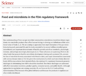
Verma MS, Finck MJ, Salmon GL, Fornelos N, Ohara TE, Ryu SH, Vlamakis H, Xavier RJ, Stappenbeck TS, Whitesides GM. ACS Chemical Biology 2018 May 18;13(5):1291-1298. doi: 10.1021/acschembio.8b00073. Epub 2018 Apr 10. New understanding of how our gut microbial communities (microbiota) transform dietary ingredients into metabolic products that affect human biology is altering our definitions of the nutritional value of foods (1, 2). We are coming to appreciate how much formation of the gut microbiota during early postnatal life and the traits encoded by its several million microbial genes (microbiome) are important determinants of healthy growth and of our metabolic, physiologic, immune, and perhaps neurologic phenotypes (3, 4). These advances are spawning efforts to develop foods that promote healthy microbiota development during early postnatal life, prevent the loss of microbial diversity associated with Western diets, and repair abnormalities associated with various disease states (5–8). But given the mechanisms by which such microbiota-directed foods (MDFs) may achieve their desired effects, the existing U.S. regulatory framework presents challenges in determining how MDFs should be classified. How these challenges are addressed will affect innovation incentives, product quality, consumer access, and public health. Although approaches to regulation vary among countries (9), we focus on the U.S. Food and Drug Administration (FDA) because of its global influence and because the products it regulates are often widely distributed. |
2017
|
Griesenauer RH, Moore, R. Kinch MS. Cell Chemical Biology. 2017 Nov 16;24(11):1315-1316. doi: 10.1016/j.chembiol.2017.11.002. A recent budget proposal from the current American administration has compelled discussions about Federal funding for the introduction of new medicines (Rear- don et al., 2017). While the funding allo- cated to the National Cancer Institute and the Office of the Director did increase from FY16 to FY17 (though no other institute changed), the President has proposed an 18% cut to funding moving forward (Achenbach and Sun, 2017; US Department of Health & Human Services, 2017). These conversations prompted an analysis of the impact of National Institutes for Health (NIH) funding for inno- vative Food and Drug Administration (FDA) drug approvals (hereafter referred to as New Molecular Entities or NMEs). |
|
Griesenauer RH, Kinch MS. Expert Review of Vaccines, 2017 Dec;16(12):1253-1266. doi: 10.1080/14760584.2017.1383159. The 2014 Ebola outbreak in West Africa, the largest outbreak on record, highlighted the need for novel approaches to therapeutics targeting Ebola virus (EBOV). Within the EBOV replication complex, the interaction between polymerase cofactor, viral protein 35 (VP35), and nucleoprotein (NP) is critical for viral RNA synthesis. We recently identified a peptide at the N-terminus of VP35 (termed NPBP) that is sufficient for interaction with NP and suppresses EBOV replication, suggesting that the NPBP binding pocket can serve as a potential drug target. Here we describe the development and validation of a sensitive high-throughput screen (HTS) using a fluorescence polarization assay. Initial hits from this HTS include the FDA-approved compound tolcapone, whose potency against EBOV infection was validated in a nonfluorescent secondary assay. High conservation of the NP-VP35 interface among filoviruses suggests that this assay has the capacity to identify pan-filoviral inhibitors for development as antivirals. |
|
Hart KM, Moeder KE, Ho CMW, Zimmerman MI, Frederick TE, Bowman GR PLoS One 2017 Jun 1;12(6):e0178678. doi: 10.1371/journal.pone.0178678. The 2014 Ebola outbreak in West Africa, the largest outbreak on record, highlighted the need for novel approaches to therapeutics targeting Ebola virus (EBOV). Within the EBOV replication complex, the interaction between polymerase cofactor, viral protein 35 (VP35), and nucleoprotein (NP) is critical for viral RNA synthesis. We recently identified a peptide at the N-terminus of VP35 (termed NPBP) that is sufficient for interaction with NP and suppresses EBOV replication, suggesting that the NPBP binding pocket can serve as a potential drug target. Here we describe the development and validation of a sensitive high-throughput screen (HTS) using a fluorescence polarization assay. Initial hits from this HTS include the FDA-approved compound tolcapone, whose potency against EBOV infection was validated in a nonfluorescent secondary assay. High conservation of the NP-VP35 interface among filoviruses suggests that this assay has the capacity to identify pan-filoviral inhibitors for development as antivirals. |
|
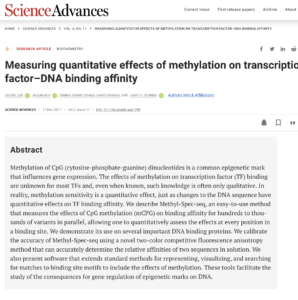
Zuo, Z, Roy B, Chang YK, Granas D, Stormo GD. Science Advances. 2017 Nov 17;3(11):eaao1799. doi: 10.1126/sciadv.aao1799 Methylation of CpG (cytosine-phosphate-guanine) dinucleotides is a common epigenetic mark that influences gene expression. The effects of methylation on transcription factor (TF) binding are unknown for most TFs and, even when known, such knowledge is often only qualitative. In reality, methylation sensitivity is a quantitative effect, just as changes to the DNA sequence have quantitative effects on TF binding affinity. We describe Methyl-Spec-seq, an easy-to-use method that measures the effects of CpG methylation (mCPG) on binding affinity for hundreds to thousands of variants in parallel, allowing one to quantitatively assess the effects at every position in a binding site. We demonstrate its use on several important DNA binding proteins. We calibrate the accuracy of Methyl-Spec-seq using a novel two-color competitive fluorescence anisotropy method that can accurately determine the relative affinities of two sequences in solution. We also present software that extends standard methods for representing, visualizing, and searching for matches to binding site motifs to include the effects of methylation. These tools facilitate the study of the consequences for gene regulation of epigenetic marks on DNA. |
|
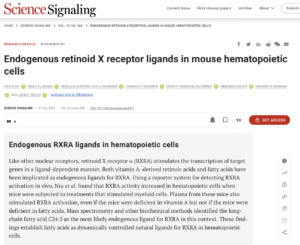
Endogenous retinoid X receptor ligands in mouse hematopoietic cells Niu H, Fujiwara H, di Martino O, Hadwiger G, Frederick TE, Menéndez-Gutiérrez MP, Ricote M, Bowman GR, Welch JS. Science Signalling. 2017 Oct 31; 10(503): eaan1011. The retinoid X receptor α (RXRA) has been implicated in diverse hematological processes. To identify natural ligands of RXRA that are present in hematopoietic cells, we adapted an upstream activation sequence-green fluorescent protein (UAS-GFP) reporter mouse to detect natural RXRA ligands in vivo. We observed reporter activity in diverse types of hematopoietic cells in vivo. Reporter activity increased during granulocyte colony-stimulating factor (G-CSF)-induced granulopoiesis and after phenylhydrazine (PHZ)-induced anemia, suggesting the presence of dynamically regulated natural RXRA ligands in hematopoietic cells. Mouse plasma activated Gal4-UAS reporter cells in vitro, and plasma from mice treated with G-CSF or PHZ recapitulated the patterns of reporter activation that we observed in vivo. Plasma from mice with dietary vitamin A deficiency only mildly reduced RXRA reporter activity, whereas plasma from mice on a fatty acid restriction diet reduced reporter activity, implicating fatty acids as plasma RXRA ligands. Through differential extraction coupled with mass spectrometry, we identified the long-chain fatty acid C24:5 as a natural RXRA ligand that was greatly increased in abundance in response to hematopoietic stress. Together, these data suggest that natural RXRA ligands are present and dynamically increased in abundance in mouse hematopoietic cells in vivo. |
|
Green JM, Barratt MJ, Kinch M, Gordon JI. Science. 2017 Jul 7;357(6346):39-40. doi: 10.1126/science.aan0836. New understanding of how our gut microbial communities (microbiota) transform dietary ingredients into metabolic products that affect human biology is altering our definitions of the nutritional value of foods (1, 2). We are coming to appreciate how much formation of the gut microbiota during early postnatal life and the traits encoded by its several million microbial genes (microbiome) are important determinants of healthy growth and of our metabolic, physiologic, immune, and perhaps neurologic phenotypes (3, 4). These advances are spawning efforts to develop foods that promote healthy microbiota development during early postnatal life, prevent the loss of microbial diversity associated with Western diets, and repair abnormalities associated with various disease states (5–8). But given the mechanisms by which such microbiota-directed foods (MDFs) may achieve their desired effects, the existing U.S. regulatory framework presents challenges in determining how MDFs should be classified. How these challenges are addressed will affect innovation incentives, product quality, consumer access, and public health. Although approaches to regulation vary among countries (9), we focus on the U.S. Food and Drug Administration (FDA) because of its global influence and because the products it regulates are often widely distributed. |
|
Liu G, Nash PJ, Johnson B, Pietzsch C, Ilagan MX, Bukreyev A, Basler CF, Bowlin TL, Moir DT, Leung DW, Amarasinghe GK. ACS Infect Dis. 2017 Mar 10;3(3):190-198. doi: 10.1021/acsinfecdis.6b00209 The 2014 Ebola outbreak in West Africa, the largest outbreak on record, highlighted the need for novel approaches to therapeutics targeting Ebola virus (EBOV). Within the EBOV replication complex, the interaction between polymerase cofactor, viral protein 35 (VP35), and nucleoprotein (NP) is critical for viral RNA synthesis. We recently identified a peptide at the N-terminus of VP35 (termed NPBP) that is sufficient for interaction with NP and suppresses EBOV replication, suggesting that the NPBP binding pocket can serve as a potential drug target. Here we describe the development and validation of a sensitive high-throughput screen (HTS) using a fluorescence polarization assay. Initial hits from this HTS include the FDA-approved compound tolcapone, whose potency against EBOV infection was validated in a nonfluorescent secondary assay. High conservation of the NP-VP35 interface among filoviruses suggests that this assay has the capacity to identify pan-filoviral inhibitors for development as antivirals. |
|
Griesenauer RH, Kinch MS. Drug Discov Today. 2017 Jul 4. pii: S1359-6446(17)30129-0. doi: 10.1016/j.drudis.2017.06.011. An overview of drugs approved by FDA in 2016 reveals dramatic disruptions in long-term trends. The number of new molecular entities (NMEs) dropped, reflecting the lowest rate of small-molecule approvals observed in almost five decades. In addition, the pace of industry consolidation slowed substantially. The impact of mergers and acquisitions decreased the total number of organizations with past approval experience and continued research and development (R&D) activities to 102, divided evenly between more established pharmaceutical and newer biotechnology companies. Despite these substantial differences, the industry continued to pursue regulatory incentives, as evidenced by a continued increase in the fraction of NMEs approved using an orphan or priority designation, and almost all oncology drugs approved in 2016 utilized these mechanisms. |

|
Kinch MS, Woodard PK. Drug Discov Today. 2017 Jul;22(7):1077-1083. doi: 10.1016/j.drudis.2017.03.006. Epub 2017 Mar 21. The posttranslational modification of proteins with poly(ADP-ribose) (PAR) regulates protein-protein interactions in DNA repair, gene expression, chrom atin structure, and cell fate determination. The PAR polymerase PARP1 binds to damaged chromatin and synthesizes PAR chains to signal DNA damage and recruit the DNA repair scaffold, XRCC1. Pharmacological blockade of PARP1 enzymatic activity impairs XRCC1-dependent repair of DNA damage and selectively kills cancer cells lacking other DNA repair functions. As such, PARP inhibitors are promising newtherapies for repair-deficient tumors such as BRCA mutated breast cancers. Although the XRCC1-PARP1 complex is relevant to the proposed therapeutic mechanism of PARP inhibitors, the physical makeup and dynamics of this complex are not well characterized at the molecular level. Here we describe a fluorescence-based, real-time assay that quantitatively monitors interactions between PARylated PARP1 and XRCC1. Using this assay, we show that the PAR posttranslational modification by itself is a high affinity ligand for XRCC1, requiring a minimum chain length of 7 ADP-ribose units in the oligo(ADP-ribose) ligand for a stable interaction with XRCC1. This discrete binding interface enables the PAR glycohydrolase (PARG) to completely disassemble the PARP1-XRCC1 complex without assistance from a mono(ADP-ribose) glycohydrolase. Our quantitative, real-time assay of PAR-dependent protein-protein interactions and PAR turnover by PARG is an excellent tool for high-throughput screening to identify pharmacological modulators of PAR metabolism that may be useful therapeutic alternatives to PARP inhibitors. |
2016
 The colonic crypt protects stem cells from microbiota-derived metabolites. The colonic crypt protects stem cells from microbiota-derived metabolites.
Kaiko GE, Ryu SH, Koues OI, Collins PL, Solnica-Krezel L, Pearce EJ, Oltz EM, Stappenbeck TS. Cell. 2016 Nov 3;167(4):1137. doi: 10.1016/j.cell.2016.10.034. In the mammalian intestine, crypts of Leiberkühn house intestinal epithelial stem/progenitor cells at their base. The mammalian intestine also harbors a diverse array of microbial metabolite compounds that potentially modulate stem/progenitor cell activity. Unbiased screening identified butyrate, a prominent bacterial metabolite, as a potent inhibitor of intestinal stem/progenitor proliferation at physiologic concentrations. During homeostasis, differentiated colonocytes metabolized butyrate likely preventing it from reaching proliferating epithelial stem/progenitor cells within the crypt. Exposure of stem/progenitor cells in vivo to butyrate through either mucosal injury or application to a naturally crypt-less host organism led to inhibition of proliferation and delayed wound repair. The mechanism of butyrate action depended on the transcription factor Foxo3. Our findings indicate that mammalian crypt architecture protects stem/progenitor cell proliferation in part through a metabolic barrier formed by differentiated colonocytes that consume butyrate and stimulate future studies on the interplay of host anatomy and microbiome metabolism. |
|
Kraft TE, Heitmeier MR, Putanko M, Edwards RL, Ilagan MG, Payne MA, Autry JM, Thomas DD, Odom AR, Cruz PW. Antimicrob Agents Chemother. 2016 Nov 21;60(12):7407-7414. Print 2016 Dec. The glucose transporter PfHT is essential to the survival of the malaria parasite Plasmodium falciparum and has been shown to be a druggable target with high potential for pharmacological intervention. Identification of compounds against novel drug targets is crucial to combating resistance against current therapeutics. Here, we describe the development of a cell-based assay system readily adaptable to high-throughput screening that directly measures compound effects on PfHT-mediated glucose transport. Intracellular glucose concentrations are detected using a genetically encoded fluorescence resonance energy transfer (FRET)-based glucose sensor. This allows assessment of the ability of small molecules to inhibit glucose uptake with high accuracy (Z′ factor of >0.8), thereby eliminating the need for radiolabeled substrates. Furthermore, we have adapted this assay to counterscreen PfHT hits against the human orthologues GLUT1, -2, -3, and -4. We report the identification of several hits after screening the Medicines for Malaria Venture (MMV) Malaria Box, a library of 400 compounds known to inhibit erythrocytic development of P. falciparum. Hit compounds were characterized by determining the half-maximal inhibitory concentration (IC50) for the uptake of radiolabeled glucose into isolated P. falciparum parasites. One of our hits, compound MMV009085, shows high potency and orthologue selectivity, thereby successfully validating our assay for antimalarial screening. |

|
Niu H, Hadwiger G, Fujiwara H, Welch JS. J Leukoc Biol. 2016 Jan 14. pii: jlb.2HI0415-146RR. PMID: 26768478. In vivo pathways of natural retinoid metabolism and elimination have not been well characterized in primary myeloid cells, even though retinoids and retinoid receptors have been strongly implicated in regulating myeloid maturation. With the use of a upstream activation sequence-GFP reporter transgene and retrovirally expressed Gal4-retinoic acid receptor α in primary mouse bone marrow cells, we identified 2 distinct enzymatic pathways used by mouse myeloid cells ex vivo to synthesize retinoic acid receptor α ligands from free vitamin A metabolites (retinyl acetate, retinol, and retinal). Bulk Kit+ bone marrow progenitor cells use diethylaminobenzaldehyde-sensitive enzymes, whereas bone marrow-derived macrophages use diethylaminobenzaldehyde-insensitive enzymes to synthesize natural retinoic acid receptor α-activating retinoids (all-trans retinoic acid). Bone marrow-derived macrophages do not express the diethylaminobenzaldehyde-sensitive enzymes Aldh1a1, Aldh1a2, or Aldh1a3 but instead, express Aldh3b1, which we found is capable of diethylaminobenzaldehyde-insensitive synthesis of all trans-retinoic acid. However, under steady-state and stimulated conditions in vivo, diverse bone marrow cells and peritoneal macrophages showed no evidence of intracellular retinoic acid receptor α-activating retinoids, despite expression of these enzymes and a vitamin A-sufficient diet, suggesting that the enzymatic conversion of retinal is not the rate-limiting step in the synthesis of intracellular retinoic acid receptor α-activating retinoids in myeloid bone marrow cells and that retinoic acid receptor α remains in an unliganded configuration during adult hematopoiesis. |
 Pathways of retinoid synthesis in mouse macrophages and bone marrow cells.
|
Kinch MS. Drug Discov Today. 2016 Jul;21(7):1046-50. doi: 10.1016/j.drudis.2016.04.008. Epub 2016 Apr 19. The myriad new molecular entities (NMEs) approved by the US Food and Drug Administration (FDA) in 2015 reflected both the opportunities and risks associated with the development of new medicines. On the one hand, the approval of 45 NMEs was among the highest ever recorded. Likewise, the diversity underlying the mechanistic basis of new medicines suggests continued broadening relative to the predominate trends of the past few decades. On the other hand, closer inspection indicates that business model decisions surrounding orphan indications and consolidation could be placing the industry in an ever-more precarious position, with severe implications for the sustainability of the entire enterprise. |

|
Patridge E, Gareiss P, Kinch MS, Hoyer D. Drug Discov Today. 2016 Feb;21(2):204-7. doi: 10.1016/j.drudis.2015.01.009. Epub 2015 Jan 21.PMID: 25617672 Natural products contribute greatly to the history and landscape of new molecular entities (NMEs). An assessment of all FDA-approved NMEs reveals that natural products and their derivatives represent over one-third of all NMEs. Nearly one-half of these are derived from mammals, one-quarter from microbes and one-quarter from plants. Since the 1930s, the total fraction of natural products has diminished, whereas semisynthetic and synthetic natural product derivatives have increased. Over time, this fraction has also become enriched with microbial natural products, which represent a significant portion of approved antibiotics, including more than two-thirds of all antibacterial NMEs. In recent years, the declining focus on natural products has impacted the pipeline of NMEs from specific classes, and this trend is likely to continue without specific investment in the pursuit of natural products. |

|
Kinch MS, Surovtseva Y, Hoyer D. Drug Discov Today. 2016 Jan;21(1):1-4. doi: 10.1016/j.drudis.2014.09.001. Epub 2014 Sep 15. Following the introduction of antibiotic therapy and widespread inoculations, cardiovascular diseases have leapt ahead of infectious diseases in terms of prevalence in much of the developed and developing world. Herein, we assess FDA-approved drugs for the treatment of cardiovascular diseases. The drug development enterprise around cardiovascular diseases has remained stable in contrast to turbulent changes in other therapeutic indications. However, upon closer inspection, the results identify narrow scope in terms of the breadth of targets and the mechanistic actions of new drugs. From the public health point of view, it is important to balance incremental change with orthogonal innovations that are needed to combat a leading cause of morbidity and mortality. |

2015

Human DNA ligase III bridges two DNA ends to promote specific intermolecular DNA end joining. Kukshal V, Kim IK, Hura GL, Tomkinson AE, Tainer JA, Ellenberger T. Nucleic Acids Res. 2015 Aug 18;43(14):7021-31. doi: 10.1093/nar/gkv652. Epub 2015 Jun 29. PMID: 26130724 Mammalian DNA ligase III (LigIII) functions in both nuclear and mitochondrial DNA metabolism. In the nucleus, LigIII has functional redundancy with DNA ligase I whereas LigIII is the only mitochondrial DNA ligase and is essential for the survival of cells dependent upon oxidative respiration. The unique LigIII zinc finger (ZnF) domain is not required for catalytic activity but senses DNA strand breaks and stimulates intermolecular ligation of two DNAs by an unknown mechanism. Consistent with this activity, LigIII acts in an alternative pathway of DNA double strand break repair that buttresses canonical non-homologous end joining (NHEJ) and is manifest in NHEJ-defective cancer cells, but how LigIII acts in joining intermolecular DNA ends versus nick ligation is unclear. To investigate how LigIII efficiently joins two DNAs, we developed a real-time, fluorescence-based assay of DNA bridging suitable for high-throughput screening. On a nicked duplex DNA substrate, the results reveal binding competition between the ZnF and the oligonucleotide/oligosaccharide-binding domain, one of three domains constituting the LigIII catalytic core. In contrast, these domains collaborate and are essential for formation of a DNA-bridging intermediate by adenylated LigIII that positions a pair of blunt-ended duplex DNAs for efficient and specific intermolecular ligation. |

Sengupta R, Barone A, Marasa J, Taylor S, Jackson E, Warrington NM, Rao S, Kim AH, Leonard JR, Piwnica-Worms D, Rubin JB. Oncotarget. 2015 Jul 30;6(21):18282-92. PMID: 26286961 Tumor growth is not solely a consequence of autonomous tumor cell properties. Rather, tumor cells act upon and are acted upon by their microenvironment. It is tumor tissue biology that ultimately determines tumor growth. Thus, we developed a compound library screen for agents that could block essential tumor-promoting effects of the glioblastoma (GBM) perivascular stem cell niche(PVN). We modeled the PVN with three-dimensional primary cultures of human brain microvascular endothelial cells in Matrigel. We previously demonstrated stimulated growth of GBM cells in this PVN model and used this to assay PVN function. We screened the Microsource Spectrum Collection library for drugs that specifically blocked PVN function, without any direct effect on GBMcells themselves. Three candidate PVN-disrupting agents, Iridin, Tigogenin and Triacetylresveratrol (TAR), were identified and evaluated in secondary in vitro screens against a panel of primary GBMisolates as well as in two different in vivo intracranial models. Iridin and TAR significantly inhibited intracranial tumor growth and prolonged survival in these mouse models. Together these data identify Iridin and TAR as drugs with novel GBM tissue disrupting effects and validate the importance of preclinical screens designed to address tumor tissue function rather than the mechanisms of autonomous tumor cell growth. |
Kim IK, Stegeman RA, Brosey CA, Ellenberger T. J Biol Chem. 2015 Feb 6;290(6):3775-83. doi: 10.1074/jbc.M114.624718. Epub 2014 Dec 4. PMID: 25477519 The posttranslational modification of proteins with poly(ADP-ribose) (PAR) regulates protein-protein interactions in DNA repair, gene expression, chrom atin structure, and cell fate determination. The PAR polymerase PARP1 binds to damaged chromatin and synthesizes PAR chains to signal DNA damage and recruit the DNA repair scaffold, XRCC1. Pharmacological blockade of PARP1 enzymatic activity impairs XRCC1-dependent repair of DNA damage and selectively kills cancer cells lacking other DNA repair functions. As such, PARP inhibitors are promising newtherapies for repair-deficient tumors such as BRCA mutated breast cancers. Although the XRCC1-PARP1 complex is relevant to the proposed therapeutic mechanism of PARP inhibitors, the physical makeup and dynamics of this complex are not well characterized at the molecular level. Here we describe a fluorescence-based, real-time assay that quantitatively monitors interactions between PARylated PARP1 and XRCC1. Using this assay, we show that the PAR posttranslational modification by itself is a high affinity ligand for XRCC1, requiring a minimum chain length of 7 ADP-ribose units in the oligo(ADP-ribose) ligand for a stable interaction with XRCC1. This discrete binding interface enables the PAR glycohydrolase (PARG) to completely disassemble the PARP1-XRCC1 complex without assistance from a mono(ADP-ribose) glycohydrolase. Our quantitative, real-time assay of PAR-dependent protein-protein interactions and PAR turnover by PARG is an excellent tool for high-throughput screening to identify pharmacological modulators of PAR metabolism that may be useful therapeutic alternatives to PARP inhibitors. |
|
Franco FM, Jones DE, Harris PK, Han Z, Wildman SA, Jarvis CM, Janetka JW. Bioorg Med Chem. 2015 May 15;23(10):2328-43. doi: 10.1016/j.bmc.2015.03.072. Epub 2015 Apr 4. PMID: 25882520 Hepatocyte growth factor activator (HGFA), matriptase and hepsin are all S1 trypsin-like serine endopeptidases. HGFA is a plasma protease while hepsin and matriptase are type II transmembrane proteases (TTSPs). Upregulated expression and activity of all three proteases is associated with aberrant cancer cell signaling through c-MET and RON tyrosine kinase cell-signaling pathways in cancer. We modeled known benzamidine protease inhibitor scaffolds into the active sites of matriptase, hepsin and HGFA to design new non-peptide inhibitors of hepsin and HGFA. First, we used a docking model of the irreversible inhibitor, Nafamostat, bound to the active site of HGFA in order to explore structure activity relationships (SAR). Compounds were screened for inhibition of HGFA activity in a kinetic enzyme assay using a chromogenic substrate. Next, we designed matched pair compound libraries of 3-amidino and 4-amidino phenylalanine (benzamidine) arginine peptidomimetics based on the structure of matriptase inhibitor, CJ-672. Compounds were screened for inhibition of HGFA, matriptase, and hepsin enzyme activity using fluorogenic substrates. Using this strategy we have discovered the first reported non-peptide small molecule inhibitors of both HGFA and hepsin. These inhibitors have differential potency and selectivity towards all three proteases. A subset of piperazinyl ureas highlighted by 25a, have excellent potency and selectivity for hepsin over matriptase and HGFA. |

2014

Monitoring Notch activation in cultured mammalian cells: luciferase complementation imaging assays. Ilagan MX, Kopan R. Methods Mol Biol. 2014;1187:155-68. doi: 10.1007/978-1-4939-1139-4_12. PMID: 25053488 Notch activation and cleavage releases the Notch intracellular domain (NICD), which translocates to the nucleus, where it associates with its DNA-binding partner CSL to recruit the coactivator MAML and additional cofactors to ultimately activate target gene expression. Taking advantage of the specific interaction between NICD and these factors, we have developed a luciferase complementation imaging (LCI)-based reporter system to quantitatively monitor Notch activation in real time in live cells. In this chapter, we describe the use of Notch LCI reporters for measuring protein interactions and performing detailed kinetic analyses of receptor activation and its responses to various stimuli. |
|
Monitoring Notch activation in cultured mammalian cells: transcriptional reporter assays. Ilagan MX, Kopan R. Methods Mol Biol. 2014;1187:143-54. doi: 10.1007/978-1-4939-1139-4_11. Review. PMID: 25053487 Upon ligand binding, Notch activation and cleavage culminates in the expression of its target genes. Hence, the use of Notch-responsive promoters to drive reporter gene expression provides a flexible and robust approach for monitoring signaling activity. In this chapter, we present an overview of Notch transcriptional reporter assays and discuss different methods for ligand-independent and ligand-dependent activation of Notch in mammalian cells. |
|
Holmes BB, Diamond MI. J Biol Chem. 2014 Jul 18;289(29):19855-61. doi: 10.1074/jbc.R114.549295. Epub 2014 May 23. Review. PMID: 24860099 Work over the past 4 years indicates that multiple proteins associated with neurodegenerative diseases, especially Tau and α-synuclein, can propagate aggregates between cells in a prion-like manner. This means that once an aggregate is formed it can escape the cell of origin, contact a connected cell, enter the cell, and induce further aggregation via templated conformational change. The prion model predicts a key role for extracellular protein aggregates in mediating progression of disease. This suggests new therapeutic approaches based on blocking neuronal uptake of protein aggregates and promoting their clearance. This will likely include therapeutic antibodies or small molecules, both of which can be developed and optimized in vitro prior to preclinical studies. |
|
T Liu Z, Chen S, Boyle S, Zhu Y, Zhang A, Piwnica-Worms DR,Ilagan MX, Kopan R. Dev Cell. 2013 Jun 24;25(6):585-98. doi: 10.1016/j.devcel.2013.05.022. PMID: 23806616 Notch2, but not Notch1, plays indispensable roles in kidney organogenesis, and Notch2 haploinsufficiency is associated with Alagille syndrome. We proposed that proximal nephron fates are regulated by a threshold that requires nearly all available free Notch intracellular domains (NICDs) but could not identify the mechanism that explains why Notch2 (N2) is more important than Notch1 (N1). By generating mice that swap their ICDs, we establish that the overall protein concentration, expression domain, or ICD amino acid composition does not account for the differential requirement of these receptors. Instead, we find that the N2 extracellular domain (NECD) increases Notch protein localization to the cell surface during kidney development and is cleaved more efficiently upon ligand binding. This context-specific asymmetry in NICD release efficiency is further enhanced by Fringe. Our results indicate that an elevated N1 surface level could compensate for the loss of N2 signal in specific cell contexts. |
|
Nickless A, Jackson E, Marasa J, Nugent P, Mercer RW, Piwnica-Worms D, You Z. Nat Med. 2014 Aug;20(8):961-6. doi: 10.1038/nm.3620. Epub 2014 Jul 27. PMID: 25064126 The year 2014 witnessed the approval by the US Food and Drug Administration (FDA) of 42 new molecular entities (NMEs), which is well above recent averages. These molecules targeted a range of molecular pathways and clinical indications, although the latter was skewed toward hepatitis C virus (HCV) infection and diabetes. By contrast, a single drug was approved for cardiovascular diseases and none for neurological indications (excepting sleeping disorders). Of note is a continued trend toward consolidation because the net number of biotechnology companies has reached its lowest point in over 25 years, raising questions about sustainability. |

|
Elhammali A, Ippolito JE, Collins L, Crowley J, Marasa J, Piwnica-Worms D. Cancer Discov. 2014 Jul;4(7):828-39. doi: 10.1158/2159-8290.CD-13-0572. Epub 2014 Apr 16. PMID: 24740997 Recently identified isocitrate dehydrogenase (IDH) mutations lead to the production of 2-hydroxyglutarate (2HG), an oncometabolite aberrantly elevated in selected cancers. We developed a facile and inexpensive fluorimetric microplate assay for the quantitation of 2HG and performed an unbiased small-molecule screen in live cells to identify compounds capable of perturbing 2HG production. Zaprinast, a phosphodiesterase 5 inhibitor, was identified as an efficacious modulator of 2HG production and confirmed to lower 2HG levels in vivo. The mechanism of action was not due to cGMP stabilization, but rather, profiling of metabolites upstream of mutant IDH1 pointed to targeted inhibition of the enzyme glutaminase (GLS). Zaprinast treatment reversed histone hypermethylation and soft-agar growth of IDH1-mutant cells, and treatment of glutamine-addicted pancreatic cancer cells reduced growth and sensitized cells to oxidative damage. Thus, Zaprinast is efficacious against glutamine metabolism and further establishes the therapeutic linkages between GLS and 2HG-mediated oncogenesis. |

2013
|
Chillakuri CR, Sheppard D, Ilagan MX, Holt LR, Abbott F, Liang S, Kopan R, Handford PA, Lea SM. Cell Rep. 2013 Nov 27;5(4):861-7. doi: 10.1016/j.celrep.2013.10.029. Epub 2013 Nov 14. PMID: 24239355 The Notch pathway is a core cell-cell signaling system in metazoan organisms with key roles in cell-fate determination, stem cell maintenance, immune system activation, and angiogenesis. Signals are initiated by extracellular interactions of the Notch receptor with Delta/Serrate/Lag-2 (DSL) ligands, whose structure is highly conserved throughout evolution. To date, no structure or activity has been associated with the extreme N termini of the ligands, even though numerous mutations in this region of Jagged-1 ligand lead to human disease. Here, we demonstrate that the N terminus of human Jagged-1 is a C2 phospholipid recognition domain that binds phospholipid bilayers in a calcium-dependent fashion. Furthermore, we show that this activity is shared by a member of the other class of Notch ligands, human Delta-like-1, and the evolutionary distant Drosophila Serrate. Targeted mutagenesis of Jagged-1 C2 domain residues implicated in calcium-dependent phospholipid binding leaves Notch interactions intact but can reduce Notch activation. These results reveal an important and previously unsuspected role for phospholipid recognition in control of this key signaling system. |

|
Boyer AP, Collier TS, Vidavsky I, Bose R. Mol Cell Proteomics. 2013 Jan;12(1):180-93. doi: 10.1074/mcp.M112.020115. Epub 2012 Oct 25. PMID: 23105007 HER2 is a receptor tyrosine kinase that is overexpressed in 20% to 30% of human breast cancers and which affects patient prognosis and survival. Treatment of HER2-positive breast cancer with the monoclonal antibody trastuzumab (Herceptin) has improved patient survival, but the development of trastuzumab resistance is a major medical problem. Many of the known mechanisms of trastuzumab resistance cause changes in protein phosphorylation patterns, and therefore quantitative proteomics was used to examine phosphotyrosine signaling networks in trastuzumab-resistant cells. The model system used in this study was two pairs of trastuzumab-sensitive and -resistant breast cancer cell lines. Using stable isotope labeling, phosphotyrosine immunoprecipitations, and online TiO(2) chromatography utilizing a dual trap configuration, ~1700 proteins were quantified. Comparing quantified proteins between the two cell line pairs showed only a small number of common protein ratio changes, demonstrating heterogeneity in phosphotyrosine signaling networks across different trastuzumab-resistant cancers. Proteins showing significant increases in resistant versus sensitive cells were subjected to a focused siRNA screen to evaluate their functional relevance to trastuzumab resistance. The screen revealed proteins related to the Src kinase pathway, such as CDCP1/Trask, embryonal Fyn substrate, and Paxillin. We also identify several novel proteins that increased trastuzumab sensitivity in resistant cells when targeted by siRNAs, including FAM83A and MAPK1. These proteins may present targets for the development of clinical diagnostics or therapeutic strategies to guide the treatment of HER2+ breast cancer patients who develop trastuzumab resistance. |
 Heparan sulfate proteoglycans mediate internalization and propagation of specific proteopathic seeds. Heparan sulfate proteoglycans mediate internalization and propagation of specific proteopathic seeds.
Holmes BB, DeVos SL, Kfoury N, Li M, Jacks, R, Yanamandra K, Ouidja MO, Brodsky FM, Marasa J, Bagchi DP, Kotzbauer PT, Miller TM, Papy-Garcia D, Diamond MI. Proc Natl Acad Sci U S A. 2013 Aug 13;110(33):E3138-47. doi: 10.1073/pnas.1301440110. Epub 2013 Jul 29. PMID: 23898162 Recent experimental evidence suggests that transcellular propagation of fibrillar protein aggregates drives the progression of neurodegenerative diseases in a prion-like manner. This phenomenon is now well described in cell and animal models and involves the release of protein aggregates into the extracellular space. Free aggregates then enter neighboring cells to seed further fibrillization. The mechanism by which aggregated extracellular proteins such as tau and α-synuclein bind and enter cells to trigger intracellular fibril formation is unknown. Prior work indicates that prion protein aggregates bind heparan sulfate proteoglycans (HSPGs) on the cell surface to transmit pathologic processes. Here, we find that tau fibril uptake also occurs via HSPG binding. This is blocked in cultured cells and primary neurons by heparin, chlorate, heparinase, and genetic knockdown of a key HSPG synthetic enzyme, Ext1. Interference with tau binding to HSPGs prevents recombinant tau fibrils from inducing intracellular aggregation and blocks transcellular aggregate propagation. In vivo, a heparin mimetic, F6, blocks neuronal uptake of stereotactically injected tau fibrils. Finally, uptake and seeding by α-synuclein fibrils, but not huntingtin fibrils, occurs by the same mechanism as tau. This work suggests a unifying mechanism of cell uptake and propagation for tauopathy and synucleinopathy. |
|
Zheng Z, Li A, Holmes BB, Marasa JC, Diamond MI. J Biol Chem. 2013 Mar 1;288(9):6063-71. doi: 10.1074/jbc.M112.413575. Epub 2013 Jan 14. PMID: 23319588 Huntington disease is a dominantly inherited neurodegenerative condition caused by polyglutamine expansion in the N terminus of the huntingtin protein (Htt). The first 17 amino acids (N17) of Htt play a key role in regulating its toxicity and aggregation. Both nuclear export and cytoplasm retention functions have been ascribed to N17. We have determined that N17 acts as a nuclear export sequence (NES) within Htt exon and when fused to yellow fluorescent protein. We have defined amino acids within N17 that constitute the nuclear export sequence (NES). Mutation of any of the conserved residues increases nuclear accumulation of Htt exon 1. Nuclear export of Htt is sensitive to leptomycin B and is reduced by knockdown of exportin 1. In HEK293 cells, NES mutations decrease overall Htt aggregation but increase the fraction of cells with nuclear inclusions. In primary cultured neurons, NES mutations increase nuclear accumulation and increase overall aggregation. This work defines a bona fide nuclear export sequence within N17 and links it to effects on protein aggregation. This may help explain the important role of N17 in controlling Htt toxicity. |

2012
|
Johnson JR, Kocher B, Barnett EM, Marasa J, Piwnica-Worms D. Bioconjug Chem. 2012 Sep 19;23(9):1783-93. Epub 2012 Aug 30. PMID: 22900707 Caspase-activatable cell-penetrating peptide (CPP) probes, designed for efficient cell uptake and specificity via cleavable intramolecular quenched-fluorophore strategies, show promise for identifying and imaging retinal ganglion cell apoptosis in vivo. However, initial cell uptake and trafficking events cannot be visualized because the probes are designed to be optically quenched in the intact state. To visualize subcellular activation events in real-time during apoptosis, a new series of matched quenched and nonquenched CPP probes were synthesized. In both native and staurosporine-differentiated RGC-5 cells, probe uptake was time- and concentration-dependent through clathrine-, caveolin-, and pinocytosis-mediated endocytic mechanisms. During apoptosis, KcapTR488, a novel dual fluorophore CPP probe, revealed by multispectral imaging a temporal coupling of endosomal release and effector caspase activation in RGC-5 cells. The novel CPPs described herein provide new tools to study spatial and temporal regulation of endosomal permeability during apoptosis. |

Lee S, Stewart S, Nagtegaal I, Luo J, Wu Y, Colditz G, Medina D, Allred DC. Cancer Res. 2012 Sep 1;72(17):4574-86. doi: 10.1158/0008-5472.CAN-12-0636. Epub 2012 Jul 2. PMID: 22751464 Molecular mechanisms mediating the progression of ductal carcinoma in situ (DCIS) to invasive breast cancer remain largely unknown. We used gene expression profiling of human DCIS (n = 53) and invasive breast cancer (n = 51) to discover uniquely expressed genes that may also regulate progression. There were 470 total differentially expressed genes (≥2-fold; P < 0.05). Elevated expression of genes involved in synthesis and organization of extracellular matrix was particularly prominent in the epithelium of invasive breast cancer. The degree of overlap of the genes with nine similar studies in the literature was determined to help prioritize their potential importance, resulting in 74 showing overlap in ≥2 studies (average 3.6 studies/gene; range 2-8 studies). Using hierarchical clustering, the 74-gene profile correctly categorized 96% of samples in this study and 94% of samples from 3 similar independent studies. To study the progression of DCIS to invasive breast cancer in vivo, we introduced human DCIS cell lines engineered to express specific genes into a “mammary intraductal DCIS” xenograft model. Progression of xenografts to invasive breast cancer was dramatically increased by suppressing four genes that were usually elevated in clinical samples of DCIS, including a protease inhibitor (CSTA) and genes involved in cell adhesion and signaling (FAT1, DST, and TMEM45A), strongly suggesting that they normally function to suppress progression. In summary, we have identified unique gene expression profiles of human DCIS and invasive breast cancer, which include novel genes regulating tumor progression. Targeting some of these genes may improve the detection, diagnosis, and therapy of DCIS. |
|
Moss BL, Elhammali A, Fowlkes T, Gross S, Vinjamoori A, Contag CH, Piwnica-Worms D. J Biol Chem. 2012 Sep 7;287(37):31359-70. doi: 10.1074/jbc.M112.364018. Epub 2012 Jul 17. PMID: 22807442 Full understanding of the biological significance of negative feedback processes requires interrogation at multiple scales as follows: in single cells, cell populations, and live animals in vivo. The transcriptionally coupled IκBα/NF-κB negative feedback loop, a pivotal regulatory node of innate immunity and inflammation, represents a model system for multiscalar reporters. Using a κB(5)→IκBα-FLuc bioluminescent reporter, we rigorously evaluated the dynamics of ΙκBα degradation and subsequent NF-κB transcriptional activity in response to diverse modes of TNFα stimulation. Modulating TNFα concentration or pulse duration yielded complex, reproducible, and differential ΙκBα dynamics in both cell populations and live single cells. Tremendous heterogeneity in the transcriptional amplitudes of individual responding cells was observed, which was greater than the heterogeneity in the transcriptional kinetics of responsive cells. Furthermore, administration of various TNFα doses in vivo generated ΙκBα dynamic profiles in the liver resembling those observed in single cells and populations of cells stimulated with TNFα pulses. This suggested that dose modulation of circulating TNFα was perceived by hepatocytes in vivo as pulses of increasing duration. Thus, a robust bioluminescent reporter strategy enabled rigorous quantitation of NF-κB/ΙκBα dynamics in both live single cells and cell populations and furthermore, revealed reproducible behaviors that informed interpretation of in vivo studies. |

Flentie K, Kocher B, Gammon ST, Novack DV, McKinney JS, Piwnica-Worms D. Cancer Discov. 2012 Jul;2(7):624-37. doi: 10.1158/2159-8290.CD-11-0201. Epub 2012 May 3. PMID: 22728436 Salmonella specifically localize to malignant tumors in vivo, a trait potentially exploitable as a delivery system for cancer therapeutics. To characterize mechanisms and genetic responses of Salmonella during interaction with living neoplastic cells, we custom-designed a promoterless transposon reporter containing bacterial luciferase. Analysis of a library containing 7,400 independent Salmonella transposon insertion mutants in coculture with melanoma or colon carcinoma cells identified five bacterial genes specifically activated by cancer cells: adiY, yohJ, STM1787, STM1791, and STM1793. Experiments linked acidic pH, a common characteristic of the tumor microenvironment, to a strong, specific, and reversible stimulus for activation of these Salmonella genes in vitro and in vivo. Indeed, a Salmonella reporter strain encoding a luciferase transgene regulated by the STM1787 promoter, which contains a tusp motif, showed tumor-induced bioluminescence in vivo. Furthermore, Salmonella expressing Shiga toxin from the STM1787 promoter provided potent and selective antitumor activity in vitro and in vivo, showing the potential for a conditional bacterial-based tumor-specific therapeutic. |
Reduced DICER1 elicits an interferon response in endometrial cancer cells. Chiappinelli KB, Haynes BC, Brent MR, Goodfellow PJ. Mol Cancer Res. 2012 Mar;10(3):316-25. doi: 10.1158/1541-7786.MCR-11-0520. Epub 2012 Jan 17. PMID: 22252463 DICER1 is essential for the generation of mature miRNAs and other short noncoding RNAs. Several lines of investigation implicate DICER1 as a tumor suppressor. Reduced DICER1 levels and changes in miRNA abundance have been associated with aggressive tumor phenotypes. The global effects of reduced DICER1 on mRNA transcript abundance in tumor cells remain largely unknown. We used short hairpin RNA to stably knock down DICER1 in endometrial cancer cell lines to begin to determine how reduced DICER1 activity contributes to tumor phenotypes. DICER1 knockdown did not affect cell proliferation but caused enhanced cell migration and growth in soft agar. miRNA and mRNA profiling in KLE cells revealed overall decreases in miRNA levels and changes in the relative abundance of many mRNAs. One of the most striking changes in mRNA levels was the upregulation of IFN-stimulated genes (ISG), the majority of which lack known miRNA target sequences. IFNβ, a key upstream regulator of the IFN response, was significantly increased in DICER1 knockdowns in the AN3CA, Ishikawa, and KLE endometrial cancer cell lines and in the normal endometrial cell line EM-E6/E7/TERT. IFNβ secreted in media from KLE and EM-E6/E7/TERT shDcr cells was sufficient to activate an IFN response in HT29 cells. The reduced miRNA processing in DICER1 knockdowns was associated with increases in pre-miRNAs in the cytoplasm. Our findings suggest that elevated pre-miRNA levels trigger the IFN response to double-stranded RNA. We thus report a novel effect of reduced DICER1 function in cancer cells. |
|
Goetzinger KR, Cahill AG, Kemna J, Odibo L, Macones GA, Odibo AO. Prenat Diagn. 2012 Oct;32(10):1002-7. doi: 10.1002/pd.3949. Epub 2012 Jul 31. PMID: 22847849 OBJECTIVE: The objective of this study was to estimate the efficiency of first-trimester a disintegrin and metalloprotease 12 (ADAM12), pregnancy-associated plasma protein A (PAPP-A), uterine artery Doppler, and maternal characteristics in the prediction of preterm birth (PTB). METHODS: This was a prospective cohort study of patients presenting for first-trimester aneuploidy screening. Maternal serum ADAM12 and PAPP-A levels were measured by immunoassay, and mean uterine artery Doppler pulsatility indices were calculated. The primary outcome was PTB <34 weeks’ gestation, and the secondary outcome was PTB <37 weeks’ gestation. Logistic regression was used to model the prediction of PTB using ADAM12, PAPP-A, uterine artery Doppler, and maternal characteristics, individually and in combination. Sensitivity, specificity, and area under the receiver-operating characteristic curves were compared between models. RESULTS: Of 578 patients, 36 (6.2%) delivered <34 weeks, and 78 (13.5%) delivered <37 weeks. For a 20% fixed false positive rate, ADAM12, PAPP-A, and uterine artery Doppler identified 58%, 52%, and 62% of patients with PTB <34 weeks and 42%, 48%, and 50% of patients with PTB <37 weeks, respectively. Combining these first-trimester parameters did not improve the predictive efficiency of the models. CONCLUSION: First-trimester ADAM12, PAPP-A, and uterine artery Doppler are each modestly predictive of PTB; however, combinations of these parameters do not further improve their screening efficiency. |

|
Zhang K, Rodriguez-Aznar E, Yabuta N, Owen RJ, Mingot JM, Nojima H, Nieto MA, Longmore GD. EMBO J. 2012 Jan 4;31(1):29-43. doi: 10.1038/emboj.2011.357. Epub 2011 Sep 27. PMID: 21952048 Snail1 is a central regulator of epithelial cell adhesion and movement in epithelial-to-mesenchymal transitions (EMTs) during embryo development; a process reactivated during cancer metastasis. While induction of Snail1 transcription precedes EMT induction, post-translational regulation of Snail1 is also critical for determining Snail1’s protein level, subcellular localization, and capacity to induce EMT. To identify novel post-translational regulators of Snail1, we developed a live cell, bioluminescence-based screen. From a human kinome RNAi screen, we have identified Lats2 kinase as a novel regulator of Snail1 protein level, subcellular localization, and thus, activity. We show that Lats2 interacts with Snail1 and directly phosphorylates Snail1 at residue T203. This occurs in the nucleus and serves to retain Snail1 in the nucleus thereby enhancing its stability. Lats2 was found to positively influence cellular EMT and tumour cell invasion, in a Snail1-dependent manner. Indeed during TGFβ-induced EMT Lats2 is activated and Snail1 phosphorylated at T203. Analysis in mouse and zebrafish embryo development confirms that Lats2 acts as a positive modulator of Snail1 protein level and potentiates its in vivo EMT activity. |

 A pharmacologic inhibitor of the protease Taspase1 effectively inhibits breast and brain tumor growth. A pharmacologic inhibitor of the protease Taspase1 effectively inhibits breast and brain tumor growth.
Chen DY, Lee Y, Van Tine BA, Searleman AC, Westergard TD, Liu H, Tu HC, Takeda S, Dong Y, Piwnica-Worms DR, Oh KJ, Korsmeyer SJ, Hermone A, Gussio R, Shoemaker RH, Cheng EH, Hsieh JJ. Cancer Res. 2012 Feb 1;72(3):736-46. doi: 10.1158/0008-5472.CAN-11-2584. Epub 2011 Dec 13. PMID: 22166309 The threonine endopeptidase Taspase1 has a critical role in cancer cell proliferation and apoptosis. In this study, we developed and evaluated small molecule inhibitors of Taspase1 as a new candidate class of therapeutic modalities. Genetic deletion of Taspase1 in the mouse produced no overt deficiencies, suggesting the possibility of a wide therapeutic index for use of Taspase1 inhibitors in cancers. We defined the peptidyl motifs recognized by Taspase1 and conducted a cell-based dual-fluorescent proteolytic screen of the National Cancer Institute diversity library to identify Taspase1 inhibitors (TASPIN). On the basis of secondary and tertiary screens the 4-[(4-arsonophenyl)methyl]phenyl] arsonic acid NSC48300 was determined to be the most specific active compound. Structure-activity relationship studies indicated a crucial role for the arsenic acid moiety in mediating Taspase1 inhibition. Additional fluorescence resonance energy transfer-based kinetic analysis characterized NSC48300 as a reversible, noncompetitive inhibitor of Taspase1 (K(i) = 4.22 μmol/L). In the MMTV-neu mouse model of breast cancer and the U251 xenograft model of brain cancer, NSC48300 produced effective tumor growth inhibition. Our results offer an initial preclinical proof-of-concept to develop TASPINs for cancer therapy. |
|
Fuentealba RA, Marasa J, Diamond MI, Piwnica-Worms D, Weihl CC. Hum Mol Genet. 2012 Feb 1;21(3):664-80. doi: 10.1093/hmg/ddr500. Epub 2011 Nov 3. PMID: 22052286 Intracellular protein aggregation is a common pathologic feature in neurodegenerative diseases such as Huntington’ disease, amyotrophic lateral sclerosis and Parkinson’ disease. Although progress towards understanding protein aggregation in vitro has been made, little of this knowledge has translated to patient therapy. Moreover, mechanisms controlling aggregate formation and catabolism in cellulo remain poorly understood. One limitation is the lack of tools to quantitatively monitor protein aggregation and disaggregation. Here, we developed a protein-aggregation reporter that uses huntingtin exon 1 containing 72 glutamines fused to the N-terminal end of firefly luciferase (httQ72-Luc). httQ72-Luc fails to aggregate unless seeded by a non-luciferase-containing polyglutamine (polyQ) protein such as Q80-cfp. Upon co-aggregation, httQ72-luc becomes insoluble and loses its enzymatic activity. Using httQ72-Luc with Q80(CFP/YFP) as seeds, we screened the Johns Hopkins Clinical Compound Library and identified leflunomide, a dihydroorotate dehydrogenase inhibitor with immunosuppressive and anti-psoriatic activities, as a novel drug that prevents polyQ aggregation. Leflunomide and its active metabolite teriflunomide inhibited protein aggregation independently of their known role in pyrimidine biosynthesis, since neither uridine treatment nor other pyrimidine biosynthesis inhibitors affected polyQ aggregation. Inducible cell line and cycloheximide-chase experiments indicate that these drugs prevent incorporation of expanded polyQ into an aggregate. This study demonstrates the usefulness of luciferase-based protein aggregate reporters for high-throughput screening applications. As current trials are under-way for teriflunomide in the treatment of multiple sclerosis, we propose that this drug be considered a possible therapeutic agent for polyQ diseases. |
|
Chinault SL, Prior JL, Kaltenbronn KM, Penly A, Weilbaecher KN, Piwnica-Worms D, Blumer KJ. Clin Cancer Res. 2012 Aug 1;18(15):4136-44. doi: 10.1158/1078-0432.CCR-12-0642. Epub 2012 Jun 12. PMID: 22693355 PURPOSE: Inhibitors of protein prenylation, including prenyltransferase inhibitors and aminobisphosphonates such as zoledronic acid, are being investigated intensively as therapeutics in cancer and other diseases. Determining whether prenylation inhibitors directly or indirectly target tumor and/or host cells is key to understanding therapeutic mechanisms. EXPERIMENTAL DESIGN: To determine which cell types can be targeted directly by distinct classes of prenylation inhibitors in vivo, we describe herein the development and implementation of a sensitive and pharmacologically specific bioluminescence-based imaging reporter that is inducible by prenylation inhibitors. RESULTS: In mouse xenograft models of breast cancer, using reporter-bearing mammary fat pad- or bone-localized tumor cells, we show that a prenyltransferase inhibitor robustly induces reporter activity in vivo. In contrast, zoledronic acid, a bone-associated aminobisphosphonate that exerts adjuvant chemotherapeutic activity in patients with breast cancer, fails to induce reporter activity in tumor cells of either model. CONCLUSIONS: Although a prenyltransferase inhibitor can directly target breast cancer cells in vivo, zoledronic acid and related aminobisphosphonates are likely to exert antitumor activity indirectly by targeting host cells. Accordingly, these findings shift attention toward the goal of determining which host cell types are targeted directly by aminobisphosphonates to exert adjuvant chemotherapeutic activity. |
2011
|
Ilagan MX, Lim S, Fulbright M, Piwnica-Worms D, Kopan R. Sci Signal. 2011 Jul 12;4(181):rs7. doi: 10.1126/scisignal.2001656. PMID: 21775282 Notch signaling regulates many cellular processes during development and adult tissue renewal. Upon ligand binding, Notch receptors undergo ectodomain shedding followed by γ-secretase-mediated release of the Notch intracellular domain (NICD), which translocates to the nucleus and associates with the DNA binding protein CSL [CBF1/RBPjκ/Su(H)/Lag1] to activate gene expression. Mammalian cells contain four Notch receptors that can have both redundant and specific activities. To monitor activation of specific Notch paralogs in live cells and in real time, we developed luciferase complementation imaging (LCI) reporters for NICD-CSL association and validated them as a specific, robust, and sensitive assay system that enables structure-function and pharmacodynamic analyses. Detailed kinetic analyses of various mechanistic aspects of Notch signaling, including nuclear translocation and inhibition of the activities of γ-secretase and ADAM metalloproteases, as well as agonist- and ligand-dependent activation, were conducted in live cells. These experiments showed that Notch-LCI is an effective approach for characterizing modulators that target Notch signaling and for studying pathway dynamics in normal and disease contexts. |

|
Srikanta D, Yang M, Williams M, Doering TL. PLoS One. 2011;6(7):e22773. doi: 10.1371/journal.pone.0022773. Epub 2011 Jul 28. PMID: 21829509 BACKGROUND: Cryptococcus neoformans causes serious disease in immunocompromised individuals, leading to over 600,000 deaths per year worldwide. Part of this impact is due to the organism’s ability to thwart what should be the mammalian hosts’ first line of defense against cryptococcal infection: internalization by macrophages. Even when C. neoformans is engulfed by host phagocytes, it can survive and replicate within them rather than being destroyed; this ability is central in cryptococcal virulence. It is therefore critical to elucidate the interactions of this facultative intracellular pathogen with phagocytic cells of its mammalian host. METHODOLOGY/PRINCIPAL FINDINGS: To accurately assess initial interactions between human phagocytic cells and fungi, we have developed a method using high-throughput microscopy to efficiently distinguish adherent and engulfed cryptococci and quantitate each population. This method offers significant advantages over currently available means of assaying host-fungal cell interactions, and remains statistically robust when implemented in an automated fashion appropriate for screening. It was used to demonstrate the sensitivity of human phagocytes to subtle changes in the cryptococcal capsule, a major virulence factor of this pathogen. CONCLUSIONS/SIGNIFICANCE: Our high-throughput method for characterizing interactions between C. neoformans and mammalian phagocytic cells offers a powerful tool for elucidating the relationship between these cell types during pathogenesis. This approach will be useful for screens of this organism and has potentially broad applications for investigating host-pathogen interactions. |

|
Gerdts J, Sasaki Y, Vohra B, Marasa J, Milbrandt J. J Biol Chem. 2011 Aug 12;286(32):28011-8. doi: 10.1074/jbc.M111.250472. Epub 2011 Jun 17. PMID: 21685387 Axon degeneration is an active, evolutionarily conserved self-destruction program by which compromised axons fragment in response to varied insults. Unlike programmed cell death, axon degeneration is poorly understood. We have combined robotic liquid handling with automated microscopy and image analysis to create a robust screening platform to measure axon degeneration in mammalian primary neuronal cultures. Using this assay, we performed an unbiased screen of 480 bioactive compounds, identifying 11 that reproducibly delay fragmentation of severed axons in vitro, including two inhibitors of glycogen synthase kinase 3 and two inhibitors of IκB kinase. Knockdown of each of these targets by shRNA lentivirus also delays axon degeneration in vitro, further supporting their role in the axon degeneration program. |


Osteopontin stimulates preneoplastic cellular proliferation through activation of the MAPK pathway. Luo X, Ruhland MK, Pazolli E, Lind AC, Stewart SA. Mol Cancer Res. 2011 Aug;9(8):1018-29. doi: 10.1158/1541-7786.MCR-10-0472. Epub 2011 Jun 14. PMID: 21673011 Alterations in the microenvironment collaborate with cell autonomous mutations during the transformation process. Indeed, cancer-associated fibroblasts and senescent fibroblasts stimulate tumorigenesis in xenograft models. Because senescent fibroblasts accumulate with age, these findings suggest that they contribute to age-related increases in tumorigenesis. Previously we showed that senescence-associated stromal-derived osteopontin contributes to preneoplastic cell growth in vitro and in xenografts, suggesting that it impacts neoplastic progression. Analysis of fibroblasts within premalignant and malignant skin lesions ranging from solar/actinic keratosis to squamous cell carcinoma revealed they express osteopontin. Given the stromal expression of osteopontin, we investigated how osteopontin impacts preneoplastic cell growth. We show that osteopontin promotes preneoplastic keratinocyte cellular proliferation and cell survival through the CD44 cell receptor and activation of the MAPK pathway. These data suggest that stromal-derived osteopontin impacts tumorigenesis by stimulating preneoplastic cell proliferation thus allowing expansion of initiated cells in early lesions. |
2010

Cardiovascular therapeutic discovery. Blaxall BC, Muslin AJ. J Cardiovasc Transl Res. 2010 Oct;3(5):429-30. doi: 10.1007/s12265-010-9217-3. Epub 2010 Aug 27. PMID: 20799008 Cardiovascular disease remains the leading cause of death in the United States and in much of the world. With obesity, diabetes, and the aging population on the rise both here and abroad, there is urgent need for development of improved cardiovascular therapeutics. |
|
Phenotypic high-throughput screening in atherosclerosis research: focus on macrophages. Muslin AJ. J Cardiovasc Transl Res. 2010 Oct;3(5):448-53. doi: 10.1007/s12265-010-9205-7. Epub 2010 Jul 13. PMID: 20625867 Atherosclerosis is a complex disease characterized by arterial lesions consisting of macrophage foam cells, smooth muscle cells, lymphocytes and other cell types. As atherosclerotic lesions mature, they can rupture and thereby trigger thrombosis that can result in tissue infarction. Macrophage foam cells develop in the subendothelial space when cells take up cholesterol from modified forms of low-density lipoprotein (LDL) and other apolipoprotein B-containing lipoproteins. Current therapies to limit atherosclerosis focus on altering the plasma lipid composition, most commonly by reducing circulating LDL levels. No current therapy is specifically designed to alter the cellular composition of atherosclerotic lesions. To address this deficit, phenotypic high-throughput drug screens have been developed to identify compounds that reduce the uptake of oxidized LDL by macrophages or to identify compounds that increase the efflux of cholesterol from macrophages. Additional phenotypic screens can be envisaged that address cellular processes in active atherosclerotic lesions including macrophage apoptosis and efferocytosis. |
|
Banerjee S, Byrd JN, Gianino SM, Harpstrite SE, Rodriguez FJ, Tuskan RG, Reilly KM, Piwnica-Worms DR, Gutmann DH. Cancer Res. 2010 Feb 15;70(4):1356-66. doi: 10.1158/0008-5472.CAN-09-2178. Epub 2010 Feb 2. PMID: 20124472 Neurofibromatosis type 1 (NF1) is a common cancer predisposition syndrome in which affected individuals develop benign and malignant nerve tumors. The NF1 gene product neurofibromin negatively regulates Ras and mammalian target of rapamycin (mTOR) signaling, prompting clinical trials to evaluate the ability of Ras and mTOR pathway inhibitors to arrest NF1-associated tumor growth. To discover other downstream targets of neurofibromin, we performed an unbiased cell-based high-throughput chemical library screen using NF1-deficient malignant peripheral nerve sheath tumor (MPNST) cells. We identified the natural product, cucurbitacin-I (JSI-124), which inhibited NF1-deficient cell growth by inducing apoptosis. We further showed that signal transducer and activator of transcription-3 (STAT3), the target of cucurbitacin-I inhibition, was hyperactivated in NF1-deficient primary astrocytes and neural stem cells, mouse glioma cells, and human MPNST cells through Ser(727) phosphorylation, leading to increased cyclin D1 expression. STAT3 was regulated in NF1-deficient cells of murine and human origin in a TORC1- and Rac1-dependent manner. Finally, cucurbitacin-I inhibited the growth of NF1-deficient MPNST cells in vivo. In summary, we used a chemical genetics approach to reveal STAT3 as a novel neurofibromin/mTOR pathway signaling molecule, define its action and regulation, and establish STAT3 as a tractable target for future NF1-associated cancer therapy studies. |
|
Honaker Y, Piwnica-Worms H. Oncogene. 2010 Jun 10;29(23):3324-34. doi: 10.1038/onc.2010.96. Epub 2010 Mar 29. PMID: 20348946 The Cdc25A protein phosphatase drives cell-cycle transitions by activating cyclin-dependent protein kinases. Failure to regulate Cdc25A leads to deregulated cell-cycle progression, bypass of cell-cycle checkpoints and genome instability. Ubiquitin-mediated proteolysis has an important role in balancing Cdc25A levels. Cdc25A contains a DS(82)G motif whose phosphorylation is targeted by beta-TrCP E3 ligase during interphase. Targeting beta-TrCP to Cdc25A requires phosphorylation of serines 79 (S79) and 82 (S82). Here, we report that casein kinase 1 alpha (CK1alpha) phosphorylates Cdc25A on both S79 and S82 in a hierarchical manner requiring prior phosphorylation of S76 by Chk1 or GSK-3beta. This facilitates beta-TrCP binding and ubiquitin-mediated proteolysis of Cdc25A throughout interphase and after exposure to genotoxic stress. The priming of Cdc25A by at least three kinases (Chk1, GSK-3beta, CK1alpha), some of which also require priming, ensures diverse extra- and intracellular signals interface with Cdc25A to precisely control cell division. |

2009
|
Saenz JB, Li J, Haslam DB. Cell Microbiol. 2010 Apr 1;12(4):516-29. doi: 10.1111/j.1462-5822.2009.01414.x. Epub 2009 Nov 27. PMID: 19951368 Infection with Shiga toxin (STx)-producing bacteria can progress to a toxemic, extraintestinal injury cascade known as haemolytic uremic syndrome (HUS), the leading cause of acute renal failure in children. Mounting evidence suggests that STx activates stress response pathways in susceptible cells and has implicated the p38 mitogen-activated protein kinase (MAPK) pathway. More importantly, some of the pathology associated with HUS is believed to be a result of a STx-induced inflammatory response. From a siRNA screen of the human kinome adapted to a high-throughput format, we found that knock-down of the MAPK-activated protein kinase 2 (MK2), a downstream target of the p38 MAPK, protected against Shiga toxicity. Further characterization of the in vitro role of MK2 revealed that STx activates the p38-MK2 stress response pathway in both p38- and MK2-dependent manners in two distinct cell lines. MK2 activation was specific to damage to the ribosome by an enzymatically active toxin and did not result from translational inhibition per se. Genetic and chemical inhibition of MK2 significantly decreased the inflammatory response to STx. These findings suggest that MK2 inhibition might play a valuable role in decreasing the immuopathological component of STx-mediated disease. |

|
Etzion Y, Hackett A, Proctor BM, Ren J, Nolan B, Ellenberger T, Muslin AJ. Circ Res. 2009 Jul 17;105(2):148-57. doi: 10.1161/CIRCRESAHA.109.195818. Epub 2009 Jun 18. PMID: 19542016 Macrophage-derived foam cells are thought to play a major role in atherosclerotic lesion formation and progression. An automated assay was established to evaluate the uptake of fluorescently labeled oxidized low-density lipoprotein (oxLDL) by a monocyte/macrophage cell line. The assay was used to screen 480 known bioactive compounds. Twenty-two active compounds were identified. Efficacy studies in peritoneal macrophages demonstrated a high rate of concordance with the initial screening results. Inhibitory compounds confirmed important previous findings and identified new drugs of interest including: 3 blockers of nuclear factor kappab activation, 2 protein kinase C inhibitors, a phospholipase C inhibitor, and 2 antipsychotic drugs. In addition, an opioid receptor agonist was found to increase the oxLDL uptake of macrophages. The involvement of nuclear factor kappaB in oxLDL uptake was validated in peritoneal macrophages in vivo. The results support a model in which oxLDL uptake is dependent on the activation of multiple intracellular signaling pathways that culminate in actin-mediated lipoprotein internalization. |
|
Naik S, Dothager RS, Marasa J, Lewis CL, Piwnica-Worms D. Clin Cancer Res. 2009 Dec 15;15(24):7529-7537. PMID: 20008853 PURPOSE: The Wnt/beta-catenin (beta-cat) signaling cascade is a key regulator of development, and dysregulation of Wnt/beta-cat contributes to selected cancers, such as colorectal, breast, and hepatocellular carcinoma, through abnormal activation of Wnt target genes. To identify novel modulators of the Wnt/beta-cat pathway that may emerge as therapeutic targets, we did an unbiased high-throughput RNA interference screen. EXPERIMENTAL DESIGN: A synthetic oligonucleotide small interfering RNA library targeting 691 known and predicted human kinases was screened in Wnt3a-stimulated human cells in a live cell luciferase assay for modulation of Wnt/beta-cat-dependent transcription. Follow-up studies of a selected high-confidence “hit” were conducted. RESULTS: A robust quartile-based statistical analysis and secondary screen yielded several kinases worthy of further investigation, including Cdc2L1, Lmtk3, Pank2, ErbB3, and, of note, vascular endothelial growth factor receptor (VEGFR)1/Flt1, a receptor tyrosine kinase (TK) with putative weak kinase activity conventionally believed to be a negative regulator of angiogenesis. A series of loss-of-function, genetic null, and VEGFR TK inhibitor assays further revealed that VEGFR1 is a positive regulator of Wnt signaling that functions in a glycogen synthase kinase-3beta (GSK3beta)-independent manner as a potential synthetic lethal target in Wnt/beta-cat-addicted colon carcinoma cells. CONCLUSIONS: This unanticipated non-endothelial link between VEGFR1 TK activity and Wnt/beta-cat signaling may refine our understanding of aberrant Wnt signaling in colon carcinoma and points to new combinatorial therapeutics targeted to the tumor cell compartment, rather than angiogenesis, in the context of colon cancer. (Clin Cancer Res 2009;15(24):7529-37). |

|
Maxwell D, Chang Q, Zhang X, Barnett EM, Piwnica-Worms D. Bioconjug Chem. 2009 Apr;20(4):702-9. doi: 10.1021/bc800516n. PMID: 19331388 Apoptosis is required for normal cellular homeostasis, and deregulation of the apoptotic process is implicated in various diseases. Previously, we developed a cell-penetrating near-infrared fluorescence (NIRF) probe based on an activatable strategy to detect apoptosis-associated caspase activity in vivo. This probe consisted of a cell-penetrating Tat peptide conjugated to an effector recognition sequence (DEVD) that was flanked by a fluorophore-quencher pair (Alexa Fluor 647 and QSY 21). Once exposed to effector caspases, the recognition sequence was cleaved, resulting in separation of the fluorophore-quencher pair and signal generation. Herein, we present biochemical analysis of a second generation probe, KcapQ, with a modified cell-penetrating peptide sequence (KKKRKV). This modification resulted in a probe that was more sensitive to effector caspase enzymes, displayed an unexpectedly higher quenching efficiency between the fluorophore-quencher pair, and was potentially less toxic to cells. Assays using recombinant caspase enzymes revealed that the probe was specific for effector caspases (caspase 3 > 7 > 6). Analysis of apoptosis in HeLa cells treated with doxorubicin showed probe activation specific to apoptotic cells. In a rat model of retinal neuronal excitotoxicity, intravitreal injection of N-methyl-d-aspartate (NMDA) induced apoptosis of retinal ganglion cells (RGCs). Eyecup and retinal flat-mount images of NMDA-pretreated animals injected intravitreally with KcapQ using a clinically applicable protocol showed specific and widely distributed cell-associated fluorescence signals compared to untreated control animals. Fluorescence microscopy images of vertical retinal sections from NMDA-pretreated animals confirmed that activated probe was predominantly localized to RGCs and colocalized with TUNEL labeling. Thus, KcapQ represents an improved effector caspase-activatable NIRF probe for enhanced noninvasive analysis of apoptosis in whole cells and live animals. |

2008
|
Pandey N, Strider J, Nolan WC, Yan SX, Galvin JE. Acta Neuropathol. 2008 Apr;115(4):479-89. doi: 10.1007/s00401-007-0332-4. Epub 2008 Jan 10. PMID: 18189141 Aggregation of amyloid-beta protein (Abeta) is a key pathogenic event in Alzheimer’s disease (AD). Curcumin, a constituent of the Indian spice Turmeric is structurally similar to Congo Red and has been demonstrated to bind Abeta amyloid and prevent further oligomerization of Abeta monomers onto growing amyloid beta-sheets. Reasoning that oligomerization kinetics and mechanism of amyloid formation are similar in Parkinson’s disease (PD) and AD, we investigated the effect of curcumin on alpha-synuclein (AS) protein aggregation. In vitro model of AS aggregation was developed by treatment of purified AS protein (wild-type) with 1 mM Fe3+ (Fenton reaction). It was observed that the addition of curcumin inhibited aggregation in a dose-dependent manner and increased AS solubility. The aggregation-inhibiting effect of curcumin was next investigated in cell culture utilizing catecholaminergic SH-SY5Y cell line. A model system was developed in which the red fluorescent protein (DsRed2) was fused with A53T mutant of AS and its aggregation examined under different concentrations of curcumin. To estimate aggregation in an unbiased manner, a protocol was developed in which the images were captured automatically through a high-throughput cell-based screening microscope. The obtained images were processed automatically for aggregates within a defined dimension of 1-6 microm. Greater than 32% decrease in mutant alpha-synuclein aggregation was observed within 48 h subsequent to curcumin addition. Our data suggest that curcumin inhibits AS oligomerization into higher molecular weight aggregates and therefore should be further explored as a potential therapeutic compound for PD and related disorders. |
